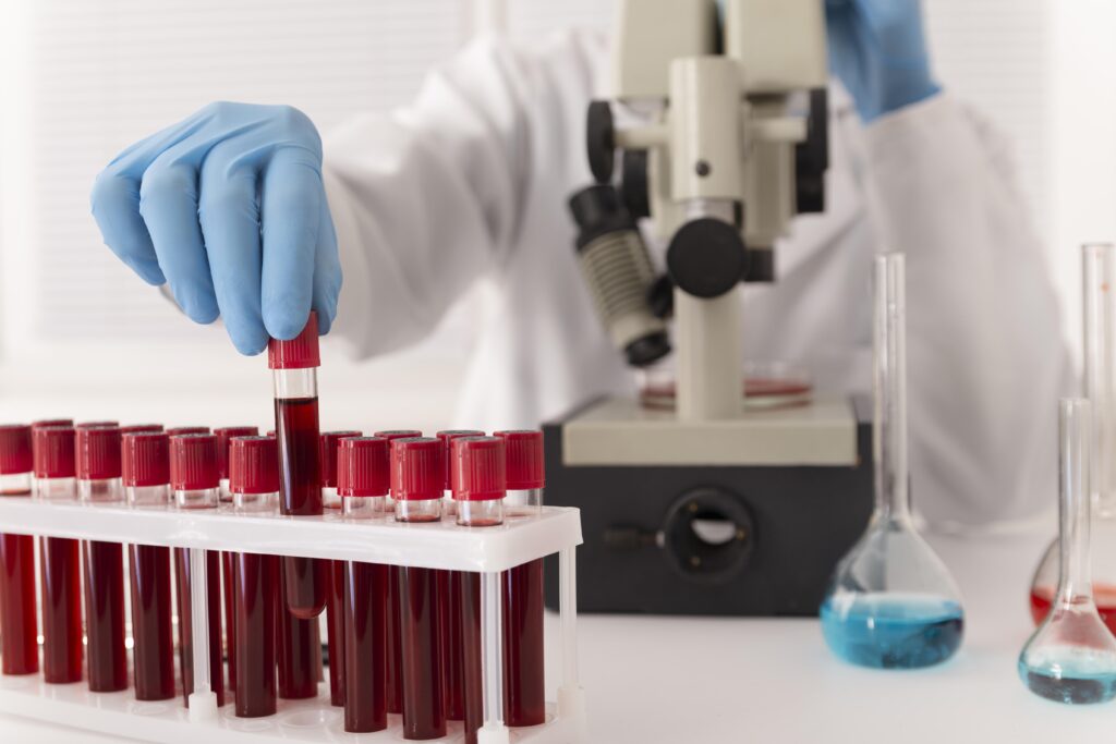
Iron overload in TDT results from frequent blood transfusions and increased gastrointestinal (GI) iron absorption due to heightened erythropoietic needs. Hepcidin, a key regulator of iron metabolism, is often disrupted, exacerbating iron accumulation. Excess iron leads to severe complications, including cardiac failure and liver cirrhosis.T2* MRI provides a non-invasive method to assess cardiac and hepatic iron levels, showing poor correlation between the two due to differing accumulation rates. The study by Soltanpour et al. also reported a high prevalence (20%) of the HFE gene mutation (H63D) in Omani patients, linked to increased GI iron absorption and serum ferritin levels.Comprehensive management includes iron chelation therapy, routine T2* MRI, and further research into genetic factors like HFE mutations to optimize care for TDT patients.